Chapter – 8
Cell: The Unit of Life
1. What is a Cell? The Cell Theory
The cell is the fundamental structural and functional unit of all living organisms. Anything less than a complete cell cannot live independently.
- Anton von Leeuwenhoek first saw and described a live cell. Robert Brown later discovered the nucleus.
- Matthias Schleiden (1838), a German botanist, observed that all plants are made of different kinds of cells.
- Theodore Schwann (1839), a British zoologist, found that animal cells have a thin outer layer (plasma membrane) and that plant cells also have a cell wall. He proposed that the bodies of animals and plants are made of cells and products of cells.
- Schleiden and Schwann together formulated the cell theory, but it did not explain how new cells are formed.
- Rudolf Virchow (1855) explained that cells divide and new cells are formed from pre-existing cells (Omnis cellula-e cellula).
Cell theory (as understood today):
- All living organisms are composed of cells and products of cells.
- All cells arise from pre-existing cells.
2. An Overview of the Cell
- Every cell has a semi-fluid matrix called cytoplasm, the main arena of cellular activities.
- Cells with a membrane-bound nucleus are eukaryotic; those without it are prokaryotic.
- Eukaryotic cells also have membrane-bound organelles such as endoplasmic reticulum, Golgi complex, lysosomes, mitochondria, microbodies and vacuoles. Prokaryotic cells lack these.
- Ribosomes are non-membrane-bound organelles found in all cells, in the cytoplasm, chloroplasts, mitochondria and on rough ER.
- Animal cells have centrosomes, which help in cell division.
Size and shape: Mycoplasma, the smallest cells, are only 0.3 µm long; bacteria are 3 to 5 µm. The largest isolated single cell is the egg of an ostrich. Human red blood cells are about 7.0 µm in diameter. Nerve cells are among the longest cells. Cells may be disc-like, polygonal, columnar, cuboid, thread-like or irregular, depending on their function.
Feature | Prokaryotic cell | Eukaryotic cell |
|---|---|---|
Nucleus | No nuclear membrane; nucleoid | Well-defined nucleus with nuclear envelope |
DNA | Single, circular, naked | Linear, with histone proteins in chromosomes |
Membrane-bound organelles | Absent | Present |
Ribosomes | 70S | 80S (70S in mitochondria and chloroplasts) |
Examples | Bacteria, blue-green algae, mycoplasma, PPLO | Protists, plants, animals, fungi |
3. Prokaryotic Cells
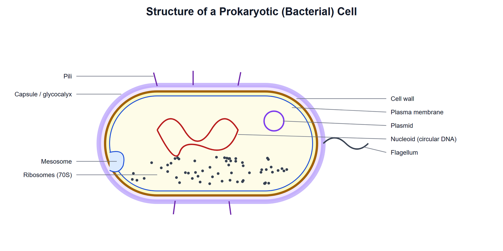
- Cell envelope: Most prokaryotic cells, especially bacteria, have a three-layered envelope: outer glycocalyx, middle cell wall and inner plasma membrane. The glycocalyx may be a loose sheath (slime layer) or thick and tough (capsule). The cell wall gives shape and prevents bursting. Based on staining by the method developed by Gram, bacteria are Gram positive (take up the stain) or Gram negative (do not).
- Mesosome: An infolding of the plasma membrane in the form of vesicles, tubules and lamellae. It helps in cell wall formation, DNA replication and distribution to daughter cells, respiration, secretion and increasing the surface area of the plasma membrane.
- Flagella: Thin filamentous extensions for motility. A bacterial flagellum has three parts: filament, hook and basal body.
- Pili and fimbriae: Pili are elongated tubular structures made of a special protein. Fimbriae are small bristle-like fibres that help bacteria attach to rocks in streams and to host tissues.
- Genetic material: Basically naked, not enveloped by a nuclear membrane. In addition to the genomic DNA (single chromosome, circular), many bacteria have small circular DNA outside the genomic DNA called plasmids. Plasmids give unique characters such as resistance to antibiotics.
- Ribosomes and inclusion bodies: Ribosomes are 70S (made of 50S and 30S subunits), about 15–20 nm. Several ribosomes attached to a single mRNA form a polysome (polyribosome). Reserve material is stored as inclusion bodies (phosphate granules, cyanophycean granules, glycogen granules). Gas vacuoles are found in blue-green and purple and green photosynthetic bacteria.
4. Eukaryotic Cells: Cell Membrane and Cell Wall
Cell membrane: Mainly composed of lipids (arranged in a bilayer) and proteins. The polar heads of lipids face outwards and the hydrophobic tails face inwards. In human RBC membranes, about 52% is protein and 40% is lipid. Proteins may be integral (partially or totally buried in the membrane) or peripheral (on the surface).
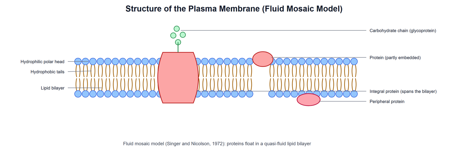
- Fluid mosaic model: Proposed by Singer and Nicolson (1972). The quasi-fluid nature of lipids allows lateral movement of proteins within the bilayer. This fluidity is important for cell growth, formation of intercellular junctions, secretion, endocytosis and cell division.
- Transport: The membrane is selectively permeable. Movement along the concentration gradient without energy is passive transport (diffusion, osmosis). Movement against the gradient using ATP is active transport (e.g. the Na+/K+ pump).
Cell wall: A non-living rigid structure outside the plasma membrane of fungi and plants. It gives shape, protects from mechanical damage and infection, helps in cell-to-cell interaction and acts as a barrier to unwanted macromolecules.
- Algae: cellulose, galactans, mannans and minerals like calcium carbonate.
- Plants: cellulose, hemicellulose, pectins and proteins.
- Primary wall is thin and capable of growth; secondary wall forms on its inner side as the cell matures.
- The middle lamella, mainly calcium pectate, holds neighbouring cells together. Plasmodesmata connect the cytoplasm of neighbouring cells.
5. The Endomembrane System
The endoplasmic reticulum, Golgi complex, lysosomes and vacuoles function in a coordinated way and are together called the endomembrane system. Mitochondria, chloroplasts and peroxisomes are not part of it.
- Endoplasmic reticulum (ER): A network of tiny tubular structures in the cytoplasm that divides the intracellular space into luminal (inside ER) and extra-luminal (cytoplasm) compartments.
- Rough ER (RER): has ribosomes on its surface; involved in protein synthesis and secretion; extensive and continuous with the outer nuclear membrane.
- Smooth ER (SER): no ribosomes; the major site for synthesis of lipids. In animal cells, lipid-like steroidal hormones are synthesised in SER.
- Golgi apparatus: First observed by Camillo Golgi (1898) near the nucleus. It consists of many flat, disc-shaped sacs (cisternae) stacked parallel to each other. It has a convex cis (forming) face and a concave trans (maturing) face. It packages materials and delivers them to intracellular targets or outside the cell. It is the important site for the formation of glycoproteins and glycolipids.
- Lysosomes: Membrane-bound vesicles formed by packaging in the Golgi apparatus. They are rich in hydrolytic enzymes (lipases, proteases, carbohydrases), which are active at acidic pH and can digest carbohydrates, proteins, lipids and nucleic acids.
- Vacuoles: Membrane-bound spaces in the cytoplasm containing water, sap, excretory products and other materials. The vacuole is bound by a single membrane called the tonoplast. In plant cells, it can occupy up to 90% of the cell volume. In Amoeba, the contractile vacuole helps in osmoregulation and excretion; in many protists, food vacuoles are formed by engulfing food particles.
6. Mitochondria, Plastids and Ribosomes

Mitochondria: Sausage-shaped or cylindrical organelles (about 0.2–1.0 µm in diameter and 1.0–4.1 µm long). Each has a double membrane: the outer membrane forms the continuous limiting boundary, and the inner membrane forms infoldings called cristae, which increase the surface area. The inner compartment is the matrix. Mitochondria are the sites of aerobic respiration and produce energy as ATP, so they are called the “power houses” of the cell. The matrix has a single circular DNA, a few RNA molecules, 70S ribosomes and components for protein synthesis. Mitochondria divide by fission.
Plastids: Found in all plant cells and in euglenoids. They have specific pigments.
- Chloroplasts: contain chlorophyll and carotenoid pigments; trap light energy for photosynthesis.
- Chromoplasts: contain fat-soluble carotenoid pigments (carotene, xanthophylls) giving yellow, orange or red colour.
- Leucoplasts: colourless plastids that store nutrients. Amyloplasts store starch (potato), elaioplasts store oils and fats, and aleuroplasts store proteins.
Chloroplasts are double-membrane bound. The space inside the inner membrane is the stroma. Flattened membranous sacs called thylakoids are arranged in stacks like piles of coins called grana. Stroma lamellae connect the thylakoids of different grana. The stroma contains enzymes for the synthesis of carbohydrates and proteins, small double-stranded circular DNA and 70S ribosomes.
Ribosomes: Granular structures first observed under the electron microscope by George Palade (1953). They are made of ribonucleic acid (RNA) and proteins and are not surrounded by any membrane. Eukaryotic ribosomes are 80S (60S + 40S subunits) and prokaryotic ribosomes are 70S (50S + 30S). “S” (Svedberg unit) stands for the sedimentation coefficient, an indirect measure of density and size.
7. Cytoskeleton, Cilia, Flagella and Centrosome
- Cytoskeleton: An elaborate network of filamentous proteinaceous structures (microtubules, microfilaments and intermediate filaments) in the cytoplasm. It helps in mechanical support, motility and maintenance of cell shape.
- Cilia and flagella: Hair-like outgrowths of the cell membrane. Cilia are small and work like oars; flagella are longer and responsible for cell movement. Both are covered by the plasma membrane. Their core, called the axoneme, has nine pairs of doublet microtubules around two central microtubules (9 + 2 array). They emerge from centriole-like structures called basal bodies. (Note: the prokaryotic flagellum is structurally different.)
- Centrosome and centrioles: The centrosome usually contains two cylindrical centrioles lying perpendicular to each other. Each centriole has a cartwheel organisation of nine evenly spaced peripheral fibrils of tubulin protein; each fibril is a triplet. Centrioles form the basal bodies of cilia and flagella and the spindle fibres that make the spindle apparatus during cell division in animal cells.
8. Nucleus and Microbodies
The nucleus was first described by Robert Brown (1831). The material of the nucleus stained by basic dyes was named chromatin by Flemming.
- Nuclear envelope: Two parallel membranes with a space between them (perinuclear space), forming a barrier between the nucleoplasm and cytoplasm. The outer membrane is usually continuous with the ER and bears ribosomes. Nuclear pores allow movement of RNA and proteins in both directions.
- Nucleoplasm: Contains the nucleolus and chromatin. The nucleolus is spherical, not membrane-bound, and is the site for active ribosomal RNA synthesis.
- Chromatin: A loose, indistinct network of nucleoprotein fibres in the interphase nucleus. It contains DNA, basic proteins called histones, some non-histone proteins and RNA. During cell division, it condenses into chromosomes. A human cell has about two metre long DNA distributed among 46 chromosomes (23 pairs).
- Chromosome structure: Every chromosome has a primary constriction, the centromere, on whose sides are disc-shaped structures called kinetochores. Some chromosomes have non-staining secondary constrictions; a small fragment beyond it is called a satellite.
Type of chromosome | Position of centromere |
|---|---|
Metacentric | Middle; two equal arms |
Sub-metacentric | Slightly away from the middle; one shorter and one longer arm |
Acrocentric | Close to one end; one extremely short and one very long arm |
Telocentric | At the terminal end |
Microbodies: Many membrane-bound minute vesicles containing various enzymes, found in both plant and animal cells (e.g. peroxisomes).
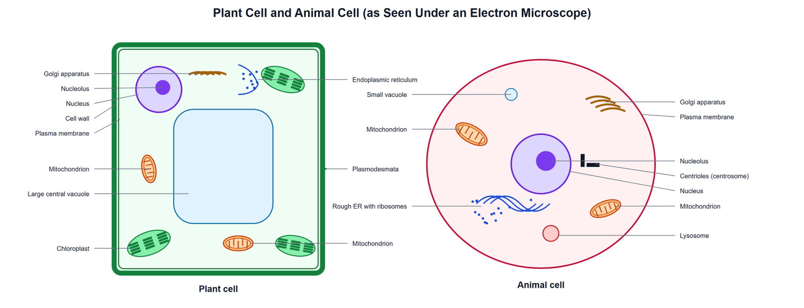
Plant cell | Animal cell |
|---|---|
Cell wall present | Cell wall absent |
Plastids present | Plastids absent |
Large central vacuole | Small vacuoles, if any |
Centrioles usually absent | Centrioles present |
Exam point: Mitochondria and chloroplasts are called semi-autonomous organelles because they have their own DNA and 70S ribosomes and can make some of their own proteins.
Conclusion: The cell is the basic unit of life. Prokaryotic cells lack a nucleus and membrane-bound organelles, while eukaryotic cells have a nucleus and many specialised organelles such as ER, Golgi apparatus, lysosomes, vacuoles, mitochondria, plastids, ribosomes, cytoskeleton and centrosomes, each with a particular role.